This study reported that:

- Chronic cocaine use in rhesus monkeys was associated with structural changes in several brain regions that mirror effects described in humans.
- Specific cocaine-induced decreases in gray matter density were associated with reduced performance on tests of memory and cognition.
- Some structural changes in brain regions involved in addiction and relapse persisted after prolonged abstinence.
Studies comparing people who use cocaine with control subjects have suggested that long-term cocaine use leads to structural changes in the brain that affect cognitive performance. However, these studies could not rule out that other, pre-existing factors may have been responsible for these effects because they did not compare the individuals’ brain structure and function before and after cocaine use. It was also unclear whether cocaine-induced changes in the brain could be reversed with abstinence.
To address these issues, Dr. Hank Jedema and colleagues from the University of Pittsburgh, VA Pittsburgh Healthcare System, and NIDA Intramural Research Program conducted repeated brain imaging and cognitive performance tests in rhesus monkeys before and after chronic cocaine self-administration. Their study demonstrated that chronic cocaine use indeed affected both brain structure and performance and that at least some of these changes persisted after prolonged abstinence.
Dr. Charles Bradberry, the study’s senior author, explains, “Having true baseline measures of both structural measures and cognitive performance helps inform and interpret clinical research into the consequences of cocaine use. Our study eliminated any potential for individual differences in vulnerability to influence the effects we attributed to cocaine exposure.”
Extended Cocaine Self-Administration Altered Gray Matter Density and Cognitive Function
The researchers trained 14 male rhesus monkeys on tests that assessed two cognitive functions—cognitive flexibility and visual working memory. They also performed a structural magnetic resonance imaging (MRI) brain scan for each animal to identify changes in gray matter density (GMD) (see Textbox). The monkeys were then assigned to a control group or to an experimental group that self-administered cocaine 4 days per week for 1 year. During that time, the team assessed the animals’ cognitive performance each week and conducted a second MRI at the end of the year. Cognitive testing continued after abstinence from cocaine, and the study concluded with a final MRI at the end of the 2-year abstinence period.
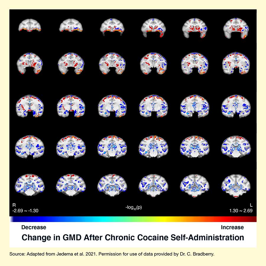
The team found that chronic cocaine use led to changes in the monkeys’ brain structure consistent with those observed in people who are dependent on cocaine. GMD decreased in several areas (e.g., the orbitofrontal, insular, and temporal cortex; amygdala; and thalamus) and increased in other areas (e.g., the caudate putamen) (see Figure 1). “We were surprised and impressed at the magnitude of the increase in GMD in the caudate putamen,” says Dr. Bradberry, adding, “These results address ambiguous clinical observations, where some studies saw a similar effect in cross-sectional comparisons, but others didn’t, and there have even been rare observations of decreased GMD.” Some of these changes (e.g., in the orbitofrontal cortex, caudate putamen, insula, and amygdala/parahippocampal cortex) were still present even after 2 years of abstinence.
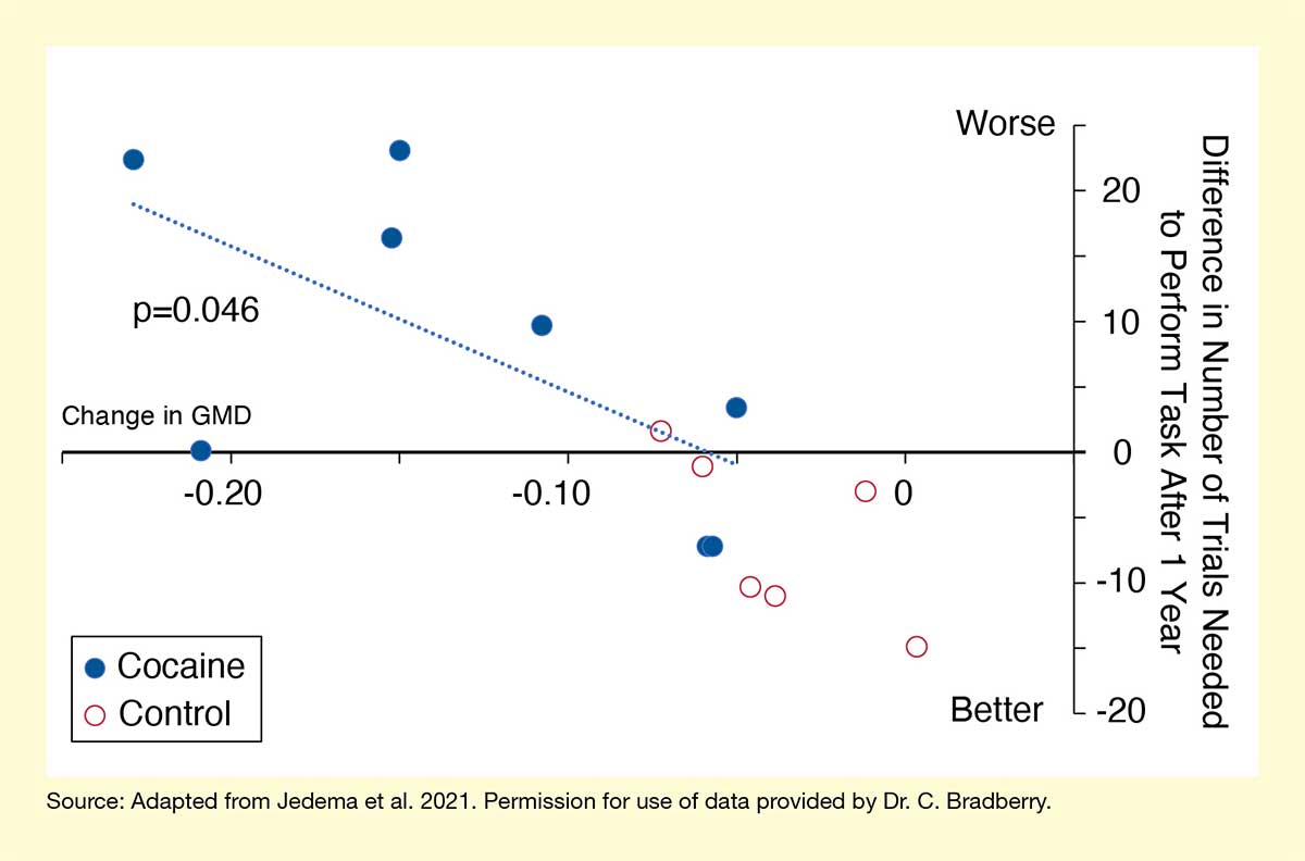
The cocaine-induced changes in some of these regions correlated with impaired cognitive performance. Thus, animals with decreased GMD in the orbitofrontal cortex and lateral parietal area had worse performance on the cognitive flexibility test (see Figure 2). Similarly, lower GMD in the insula and temporal cortex was associated with reduced performance in the visual working memory test.
Dr. Jedema and colleagues concluded that chronic cocaine use causes specific changes in brain structure that influence cognitive performance and can persist even after prolonged abstinence. Studies comparing people with and without cocaine use disorder detected similar patterns. The researchers are currently analyzing brain tissues from some of the animals to identify potential cellular mechanisms that could be driving the cocaine-induced structural changes, such as by comparing areas that showed decreased GMD with those with increased GMD.
The findings may have additional implications. The team noted that some of the persistent structural changes occurred in brain regions thought to be involved in response inhibition and addiction in humans. Thus, it is possible that these brain alterations may be a factor contributing to relapse risk.
This study was supported by NIDA grant DA025636.
Gray Matter Density
The brain’s gray matter comprises the cell bodies of the neurons in the brain and is largely responsible for muscle control, sensory perception, and various cognitive functions. Numerous studies have established that greater gray matter volume is associated with better cognitive performance. However, gray matter density also influences cognitive performance. Gray matter density refers to the proportion of matter within a given brain area that is gray matter (i.e., nerve cell bodies) rather than white matter (i.e., nerve cell fibers) or fluid, as determined during brain imaging. Both gray matter volume and gray matter density can change over the course of the life span and in response to environmental influences, such as drug use. Decreases in both gray matter volume and gray matter density in certain brain regions have been associated with reduced cognitive performance on specific tasks.
- Text Description of Figure 1
-
The figure shows five rows of six slices through the brain of Rhesus monkeys. In each slice, colored areas indicate brain regions where the GMD changed after chronic cocaine self-administration compared with baseline. At the bottom, a color scale indicates the degree of change. It ranges from dark blue on the left, indicating a decrease in GMD, through light blue, green, yellow, orange, and red to dark red on the right, indicating an increase in GMD.
- Text Description of Figure 2
-
The figure shows the relationship between structural changes in the orbitofrontal cortex of Rhesus monkeys and cognitive flexibility. The horizontal x-axis indicates the changes in GMD on a scale from 0 on the right to -0.20 on the left. The vertical y-axis on the right indicates the difference in the number of trials the animals needed to perform a task assessing cognitive flexibility after 1 year of cocaine self-administration, on a scale from -20 (indicating better performance) to +20 (indicating worse performance). Filled blue circles represent animals that self-administered cocaine; open red circles represent control animals. A dotted blue line sloping from the x-axis upwards to the top left indicates the relationship between greater reductions in GMD after cocaine self-administration and worse cognitive performance.
Source
- Jedema, H.P., Song, X., Aizenstein, H.J., et al. Long-term cocaine self-administration produces structural brain changes that correlate with altered cognition. Biological Psychiatry. 89(4):376-385, 2021. doi: 10.1016/j.biopsych.2020.08.008.