
![]()
Köhler disease
Description
Köhler disease is a rare, idiopathic self-limiting osteochondrosis caused by avascular necrosis of the tarsal navicular bone presenting in young children with dorsomedial foot pain and limp
Most common in young children (usually 3-9 yrs); 80% of cases are in males; and unilateral in 75% of cases. Most likely caused by excessive strain on the tarsal navicular bone and its associated blood vessels before the bone is completely ossified.
Signs and symptoms include swelling and redness to the dorsomedial midfoot; tenderness along the length of the arch; limp or abnormal gait.
Treatment is usually nonoperative with NSAIDs, and a short period of cast immobilisation.
Kohler disease is distinct from Müller-Weiss disease, a complex idiopathic foot condition with deformity of the tarsal navicular presenting in adulthood
Case example
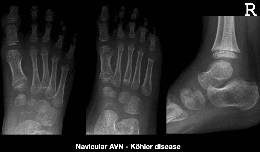
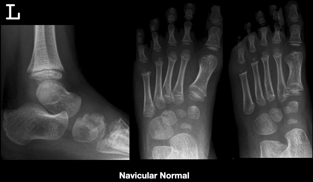
Below are images of right foot Navicular avascular necrosis (Köhler disease) in a 5 year old male


Spontaneous adult onset osteonecrosis of the tarsal navicular is known as Mueller-Weiss syndrome (also Brailsford disease)
History of Köhler disease
1908 – First described by German radiologist Alban Köhler (1874-1947) in his MWW article Uber eine häufige, bisher anscheinend unbekannte Erkrankung einzelner kindlicher Knochen [About a common, so far apparently unknown disease of individual children’s bones]

Original
English
Es handelte sich um Knaben im Alter von 5-9 Jahren (..) Alle drei Patienten klagten über mehr oder weniger heftige Schmerzen in der Gegend des Os naviculare (…) Die Röntgenuntersuchung ergab einen recht eigenartigen (…) Befund. Ueberall zeigte sich das Os naviculare in die Augen fallend erkrankt, während alle anderen Knochen des Fusses einen normalen Anblick boten. Das Navikulare war in vierfacher Beziehung verändert, und zwar in seiner Grösse, seiner Gestalt, seiner Architektur und seinem Kalkgehalt.
Der Krankheitsverlauf zog sich auf 2-3 Jahre hin (…) es erfolgte Heilung (…) nicht nur im klinischen Sinne, sondern wie die Röntgenbilder anzunehmen zwingen, auch in anatomischem Sinne. Die Prognose des Leidens ist also durchaus günstig zu stellen.
In question were young lads aged 5-9 years old. All patients complained of more or less severe pains in the region of the navicular bone. The Röntgen examination demonstrated a most peculiar finding. Everywhere the navicular bone appeared diseased, whilst all other bones of the foot presented normally. The navicular was changed in four aspects, namely its size, shape, architecture, and calcification.
The disease took its course in 2-3 years (…) healing was evident(…) not only in the clinical sense, but also in the anatomical sense, as the X-rays force us to acknowledge. The prognosis of this suffering is therefore quite favourable.
Associated Persons
- Alban Köhler (1874-1947)
- Walther Müller (1888-1949)
- Konrad Weiss (1891-1976)
- James F Brailsford (1888-1961)
Alternative names
- Kohler disease, Koehler disease
- Osteonecrosis; Osteochondrosis; Osteochondritis of the tarsal navicular
References
Original articles
- Köhler A. Uber eine häufige, bisher anscheinend unbekannte Erkrankung einzelner kindlicher Knochen. Munchener medizinische Wochenschrift, 1908; 55: 1923-1925
Review articles
- Williams GA, Cowell HR. Kohler’s disease of the tarsal navicular. Clin Orthop Relat Res. 1981; 158: 53-8.
- Ippolito E, Ricciardi Pollini PT, Falez’ F. Köhler’s disease of the tarsal navicular: long-term follow-up of 12 cases. J Pediatr Orthop. 1984; 4(4): 416-7
- Shastri N, Olson L, Fowler M. Kohler’s Disease. West J Emerg Med. 2012; 13(1): 119–120.
- Gomez A, Cadogan M. Eponymythology of foot injuries. LITFL
- Cadogan M. Köhler disease. Eponym A Day. Instagram
[cite]
eponymictionary
the names behind the name

Resident medical officer in emergency medicine MB ChB (Uni. Dundee) MRCS Ed. Avid traveller, yoga teacher, polylinguist with a passion for discovering cultures.