Brain scans could spot signs of Alzheimer’s years before symptoms begin, according to new research. Increased volume of a region called the choroid plexus is linked to greater cognitive impairment, say scientists.
The discovery offers hope of a Alzheimer’s disease screening program to identify vulnerable individuals. Detecting early changes is vital to developing treatments. Drugs trials have failed to date because they are given to patients after the disease has taken hold.
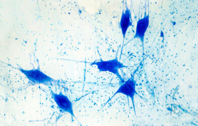
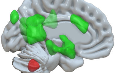
The choroid plexus is a network of blood vessels, connective tissue and cells found in spaces called ventricles. It acts as a gateway for immune cells from the blood and also produces cerebrospinal fluid, a clear liquid that surrounds the brain and spinal cord each day. This washes away waste products and rogue proteins called amyloid and tau that kill neurons.
“Researchers believe impaired clearance rather than overproduction of abnormal amyloid and tau is responsible for Alzheimer’s disease,” says senior author Won-Jin Moon, of Konkuk University in Seoul, Korea, in a statement per South West News Service. “Thus, we assume the abnormal status of choroid plexus is linked to the failure of clearance leading to waste and toxic protein accumulation in the brain and failure of immune surveillance leading to neuroinflammation.”
The findings are based on MRI (magnetic resonance imaging) scans of 532 people at various stages of cognitive impairment. Choroid plexus mass was greater in those with Alzheimer’s, with bigger size correlating with worsening memory. It also had negative effects on executive function, the wide-ranging set of mental skills governing things like self-control and planning.
Previously, little was known about the choroid plexus’ imaging profile in cognitive impairment.
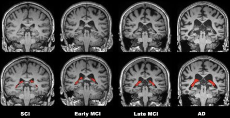
“Our study found the enlarged choroid plexus volume is independently associated with increased cognitive impairment,” says Moon. “We found no relationship between choroid plexus volume and amyloid pathology but a clear relationship between the choroid plexus volume and cognitive impairment severity.”
The results point to new possibilities for MRI’s role in the diagnosis of Alzheimer’s.
“I think our findings on the choroid plexus can suggest it as a new potential imaging surrogate for an impaired clearance system and neuro-inflammation,” Moon adds.
Other potential clinical applications include helping researchers develop new target drugs or treatments. Eventually, choroid plexus measurements could help speed therapy to those who need it most.
“If we combine choroid plexus volume and hippocampal volume in a screening stage, it may help us better discriminate the more vulnerable patients from the less vulnerable ones,” says Moon.
The researchers plan a follow-up study that will track patients over a long period of time. They will explore changes in choroid plexus volume as the disease progresses.
The number of dementia cases worldwide will triple to more than 150 million in the next three decades, according to a recent study. Current drugs can treat the symptoms, but not the cause.
The study is published in the journal Radiology.
Report by South West News Service writer Mark Waghorn.